July 15, 2020
When the heart recovers from injury, the blood flowing through its vessels is essential. But lymph – the colourless fluid that circulates in a parallel network – and the lymphatic vessels in which it moves are just as crucial, according to researchers at the Weizmann Institute of Science.
The researchers showed that without an adequate network of lymphatic vessels, the hearts of zebrafish – which can completely regenerate following injury – fail to heal, even in the presence of blood vessels.
The researchers also identified two distinct types of lymphatic vessels in the fish heart, which form by different mechanisms and perform different tasks. These findings may help develop new ways for promoting heart repair and facilitating the growth of organs for transplant.
The body’s network of lymphatic vessels serves as a conduit for immune cells and drains fluid, as well as providing individual services to various organs. Professor Karina Yaniv of Biological Regulation Department and her team wanted to learn what role the lymphatic vessels play in the heart, particularly following injury.
Zebrafish are perfect for exploring this question because unlike mammals, they have a remarkable capacity for heart regeneration: Within approximately two months after being injured, up to one-third of their heart tissue can regrow without scarring.
An injured mammalian heart, in contrast, does not naturally regenerate at all, instead forming scar tissue at the injury site.
In earlier research, Yaniv and colleagues had shown that in the zebrafish embryo, lymphatic vessels form by two separate mechanisms: either by sprouting and growing from pre-existing vessels, or by differentiating from progenitor cells called angioblasts.
In the new study, the researchers found that in adult zebrafish, these two mechanisms are recapitulated. Moreover, each set of lymphatic vessels features its own genetic markers and responds to its own molecular signals. The scientists found that both distinct types of lymphatic vessels are also found in the hearts of mice, indicating that the disparate mechanisms by which they develop have been conserved in the course of evolution.
The researchers then asked whether either or both types play a role in regeneration. After injuring the hearts of zebrafish, Yaniv and her group discovered that the hearts’ regenerating area contained lymphatic vessels of just one type – those derived from isolated lymphatic cells, rather than vessels. These cells then merged together into vessels. Although these vessels eventually connected with the larger lymphatic vessels that belong to the other type, they remained distinct. Thus, the experiments revealed that the two types of lymphatic vessels perform different functions in the heart: It is evidently mainly the progenitor cell-derived ones that contribute to heart regeneration after injury.
The researchers then showed that these lymphatic cells do more than facilitate heart regeneration – the process cannot take place without them.
When the scientists studied mutant zebrafish with no lymphatic vessels at all, the hearts of these mutants failed to regenerate after injury even though their blood vessels were entirely normal.
“We’ve shown that the lymphatic system plays a crucial role in heart regeneration in the zebrafish,” said Professor Yaniv.
“By clarifying this role further, we may learn what the fish heart ‘knows’ about regeneration that the mammalian one doesn’t, and use this knowledge to heal human hearts.”
A more detailed understanding of this role may indeed help develop new ways of preventing scarring and promoting healing after heart attacks or other types of injury to the heart muscle. In addition, knowing exactly how the lymphatic system contributes to heart muscle growth may in the future help to grow heart tissue in laboratory conditions for the purposes of transplantation.
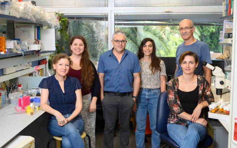
Study authors included Dr Dana Gancz, Gal Perlmoter, Dr Jonathan Semo, Hila Raviv and Noga Moshe of Biological Regulation Department; Brian C Raftrey and Professor Kristy Red-Horse of Stanford University; Dr Rubén Marín-Juez, Professor Ryota L. Matsuoka and Professor Didier Y. R. Stainier of the Max Planck Institute for Heart and Lung Research, Bad Nauheim, Germany; Dr Ravi Karra and Professor Kenneth D. Poss of Duke University; and Dr Yoseph Addadi and Ofra Golani of Weizmann’s Life Sciences Cored Facilities Department.
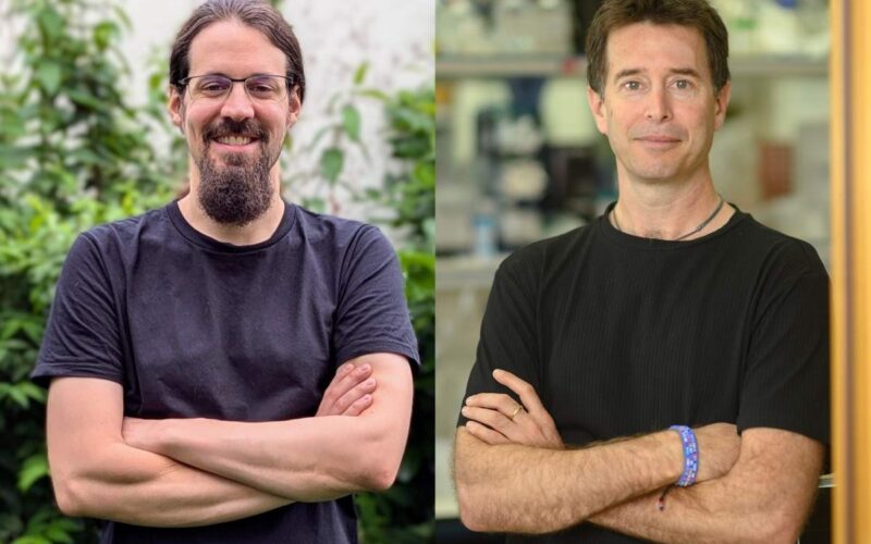
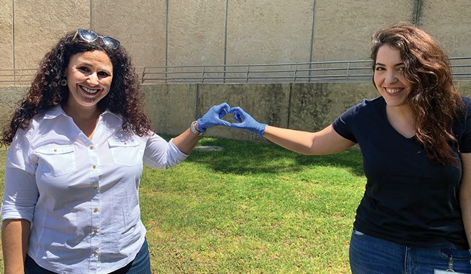
Professor Karina Yaniv and Gal Perlmoter

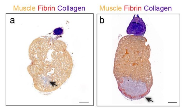
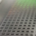
Cross-section of a zebra fish heart 30 days after injury. The injured spot (arrow) regenerates without forming a scar in a fish that has intact lymphatic vessels (left), but not in a mutant fish lacking such vessels (right)
Tagged: blood flow, heat, injury, Lymphatic vesels, vessels, Weizmann Institute of Science, Yaniv, zebrafish