February 5, 2016
Proteins are the molecular machines that make living things function. Understanding, measuring and visualising their structural changes as they perform biological functions are therefore imperative. However to see such changes, proteins must be studied not in a single state, as found in crystals, but free moving in solution, which is their natural environment. Yet this poses a major challenge.
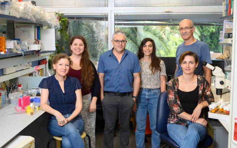
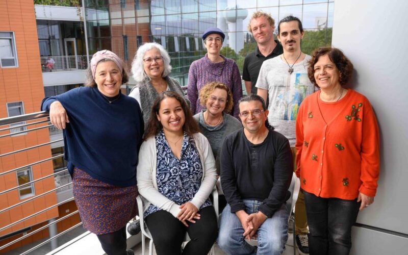
This challenge was the focus of collaborative research between the Australian National University (ANU), Australia and the Weizmann Institute of Science (WIS), Israel – work that has provided key tools for not only ‘seeing’ proteins but may ultimately help scientists further understand how drugs target cells in the body.
As atoms and molecules are so small, to see these proteins at work is very difficult. In the approach chosen by the WIS-ANU collaboration, researchers tag them with specific molecules that in turn can be seen using electron paramagnetic resonance (EPR) spectroscopy, which is the best method available to accurately measure the distance between tags on a nanoscale and the variability of these distances in solutions. Such measurements allow researchers to track changes in the protein structure as it interacts with other proteins or chemicals interfering with its function.
Funded by a two year Weizmann Australia grant *, the ANU and WIS research groups combined ANU’s cutting-edge expertise in tagging proteins with suitable molecules, with WIS’s experience in measuring the distance between them using EPR. The Weizmann Institute possesses one of the most sensitive high field EPR spectrometers in the World.
The final research results included the development of two new molecular tags, which provided important information on the molecular motions of a glutamate-binding protein and of the calcium-binding protein calmodulin.
Glutamate is a key neurotransmitter in the brain and the glutamate binding protein, which is used by several scientific groups to develop glutamate biosensors, was the first the researchers studied. Using protein from common bacteria they sought to see how the protein changes its structure to bind glutamate, something not yet known. To do this the ANU laboratory developed a new labelling method using two man-made amino acids which were introduced into the protein at specific sites, informed by a known atomic-resolution structure of the protein state with bound glutamate. A variety of 15 tag combinations were then made and sent to WIS in Israel for measurement. High quality measurements resulted and showed the structure of the tagged protein was indeed similar to the untagged structure and proved that this new labelling scheme provided good results. The work resulted in publications printed in Chemical Communications[i] and accepted in the Journal of Biomolecular NMR.[ii]
The second protein studied – calmodulin – is a calcium binding protein which goes through large structural changes, and which the scientists studied using a previously established tag. These are currently being measured by WIS but preliminary results indicate that this binding protein operates very differently to the glutamate binding protein, with large structural changes occurring upon binding of calcium. These results should provide important information about the various ways proteins operate.
Finally the rigid tag devised by ANU in collaboration with the group of Dr Bim Graham at Monash University to label cysteine – called a gadolinium tag – was measured by WIS. It showed it could be very useful for high sensitivity measurements and precise distance measurements, therefore providing an important tool for performing accurate distance measurements for more complex probing and understanding of proteins in the future. This work has also been accepted for publication in the Journal of Physical Chemistry Letters.[iii]
During the course of the research project ANU’s Professor Gottfried Otting spent a month at WIS in January 2014 and WIS’ Professor Daniella Goldfarb visited ANU in December 2014 where she delivered a three day EPR course at the University.
‘Seeing’ protein molecules at work is an age-old dream of biochemists and biophysicists and this new collaborative work has significantly improved this process. The researchers expect the technological tools developed in this project will set the stage for more detailed structural and motional studies of proteins that cannot be addressed by any other means.
To read the detailed scientific report click the Goldfarb & Otting Final Report.
Professors Otting and Goldfarb discuss their work
[i] Abdelkader, EH; Feintuch, A; Yao, X; Adams, LA; Aurelio, L; Graham B; Goldfarb, D; Otting, G: Protein conformation by EPR spectroscopy using lanthanide tagging of genetically encoded amino acids. Chem. Commun. 2015. 51, 15898-15901.
[ii] Abdelkader, EH; Feintuch, A; Yao, X; Adams, LA; Aurelio, L; Graham B; Goldfarb, D; Otting, G: Pulse EPR-enabled interpretation of scarce pseudocontact shifts induced by lanthanide binding tags. J. Biomol. NMR 2015, in press.
[iii] Abdelkader, EH; Lee, MD; Feintuch, A; Ramirez Cohen, M; Swarbrick, JD; Otting, G; Graham, B; Goldfarb, D: A new Gd3+ spin label for Gd3+ – Gd3+ distance measurements in proteins produces narrow distance distributions. J.Phys.Chem.Lett. 2015 in press.
* These grants were funded by a group of Australian donors. We thank them for their generous and visionary support of this program.


