A mysterious menace lurks in the oceans – threatening algal blooms. Now, Weizmann Institute researchers have developed a new way to track the culprit – giant viruses – and identify their traces in specific types of tiny algae that they attack.
They were said to come from outer space, and there were even claims that they were actually bacteria and that they undermined the very definition of viruses.
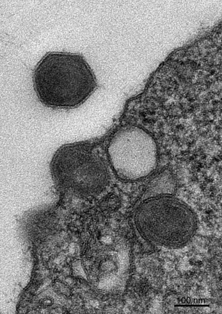
Giant viruses, nicknamed ‘giruses’, contain enormous quantities of genetic material – up to 100 times more than other viruses – and some are larger than certain bacteria. They were practically unknown to science until the early 21st century, yet discoveries made over the past two decades suggest that they can have a major impact on life on Earth.
Giant viruses inhabiting the oceans infect, among others, various species of single-celled algae, photosynthetic organisms that are responsible for about half of the Earth’s oxygen production and around half of the global carbon fixation. Viral infection can cause a rapid collapse of algal blooms – accumulations of algae stretching across tens of thousands of kilometres in the ocean – and this can, in turn, substantially affect extensive marine, atmospheric and terrestrial ecosystems. However, we still know very little about the natural hosts of giant viruses, that is, which species of algae are infected by each type of virus.
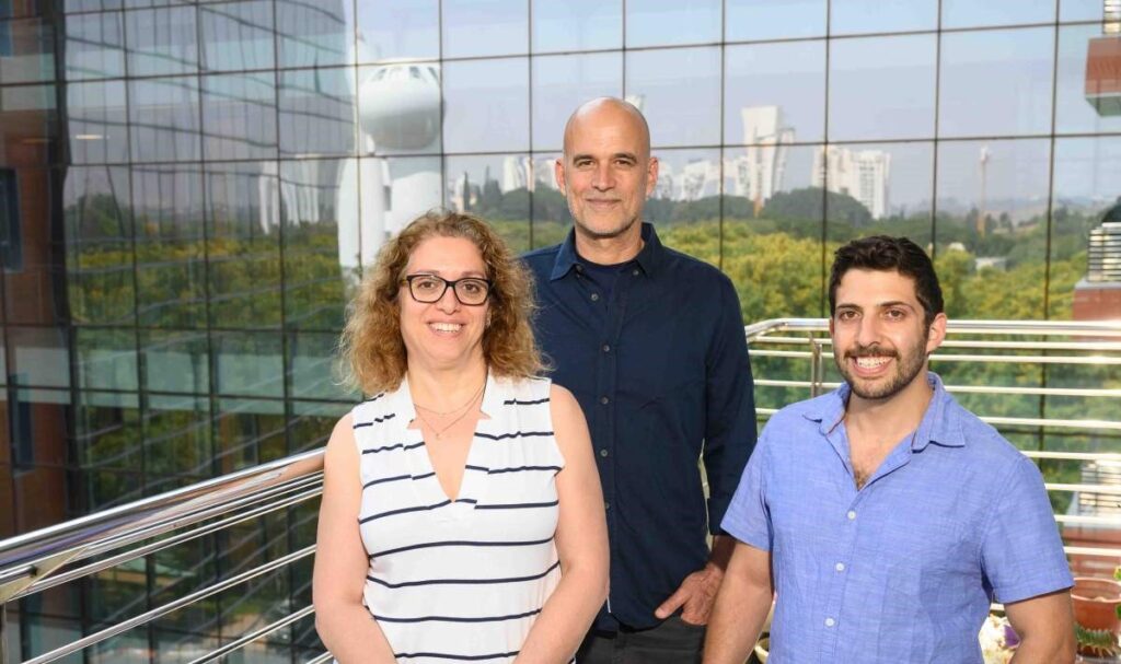
In a new study, researchers from Professor Assaf Vardi’s lab in the Plant and Environmental Sciences Department at the Weizmann Institute of Science used single-cell RNA sequencing to analyse samples collected from an algal bloom in the fjords of Norway. This enabled them to map out, in unprecedented detail, the relationships between giant viruses and the single-celled marine organisms, including algae, that each of them infects.
Until recently, the most effective way of analysing the populations of viruses and algae in the ocean was to examine them in bulk, that is, analyse all of the genetic material isolated from ocean water samples that were swarming with viruses and single-celled marine organisms.
“These studies advanced our knowledge about the distribution of algae and viruses in the water, but much information was still lacking,” Vardi explained.
“We were able to learn which species were abundant in a particular sample but not to identify active infection, so we were limited in our ability to measure the impact of viruses on algal bloom or collapse. Likewise, we couldn’t identify populations that were less prevalent in the sample, determine the natural host of each virus or learn whether the viruses were active in the cells.”
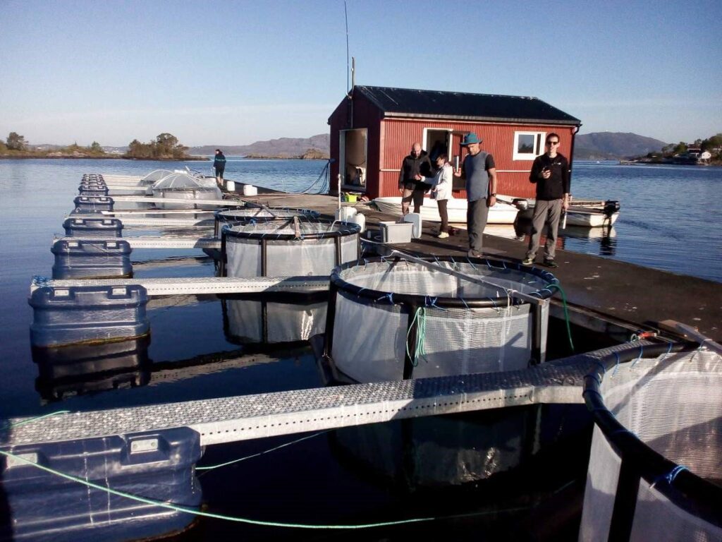
To overcome these limitations and to examine the interactions that exist in the natural environment, the researchers travelled to the Norwegian fjords, where they induced an algal bloom by adding nutrients to the water and by mimicking the conditions that lead to blooms. Back in the laboratory, they sequenced the RNA of individual cells in their samples.
These RNA molecules indicate which genes are active in the organisms sampled at any given time. In contrast to regular RNA sequencing, in which all of the RNA from all the organisms in the sample is pulled together, the researchers used single-cell RNA sequencing. This advanced, high-resolution method allows them to tag individual cells and identify which RNA uniquely appears in that particular cell.
Using this method, the researchers were able to identify the active genes in the cells and, by comparing them to existing databases, work out to which species each cell in the sample belonged. They were not satisfied, however, with just mapping the hosts; they also wanted to establish which viruses infect each cell in the natural environment.
“We realized that by meticulously analysing the RNA, we could produce two kinds of data, not only identifying the species of algae but also determining whether the algae had been infected by a giant virus and, if so, which one,” explained Amir Fromm, the PhD student from Professor Vardi’s lab who led the study.
Prior to this study, there was no database classifying giant viruses that infect algae, so the researchers approached Professor Frank Aylward, an expert on the evolution of viruses at Virginia Tech, who, with his own team, put together a unique database of giant viruses, based on the RNA extracted from the cells.
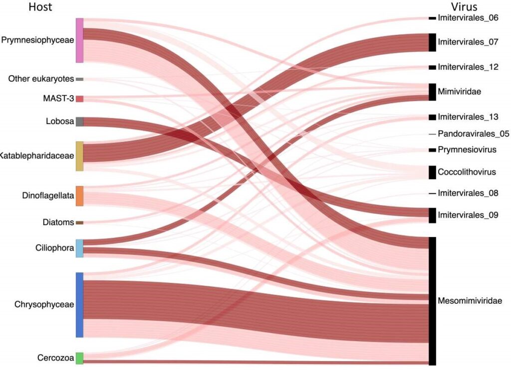
Of the tens of thousands of cells that were examined, many belonged to the dominant algae in that bloom, Emiliania huxleyi. In an earlier study, Vardi and his team had already identified the specific giant virus that infects this alga. This time, they focused on other virus-host couples, present in tiny quantities in the samples.
In total, they found 972 infected cells, of which 71 did not belong to the dominant algae in the bloom and which included several giant virus-host combinations that were previously unknown.
For example, they discovered that the Imitervirales-07 virus infects algae cells from the Katablepharidaceae family – a connection that had not previously been described.
When they examined the RNA of the virus in these cells, they found that it produces proteins that have a direct impact on the fate of the cells, including proteins capable of causing the cells to commit suicide (programmed cell death) during the late phase of infection. The researchers even found a smoking gun: They demonstrated a link between the appearance of the virus in the examined samples and the collapse of the host algae population shortly afterward.
Since the researchers managed to successfully tackle the computational challenge of characterising infected cells in samples from tiny amounts of RNA, they believe that their method can also be used to identify interactions of algae with viruses and other pathogens in other parts of the world, including extreme climate environments such as the poles and Alpine lakes.
“Because of processes linked to climate change, ancient pathogens are being released from melting icecaps at the poles. This new method will allow us to identify these pathogens with much greater ease and to help humanity prepare for the environmental threat that they may pose,” Vardi said.
This study was part of collaborative research that included Dr Gur Hevroni, Dr Flora Vincent and Dr Daniella Schatz from Vardi’s laboratory at the Weizmann Institute; and Dr Carolina Martinez-Gutierrez from Aylward’s laboratory at Virginia Tech.